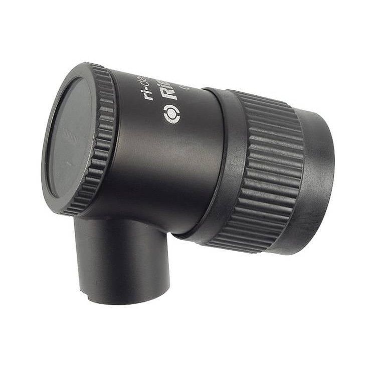
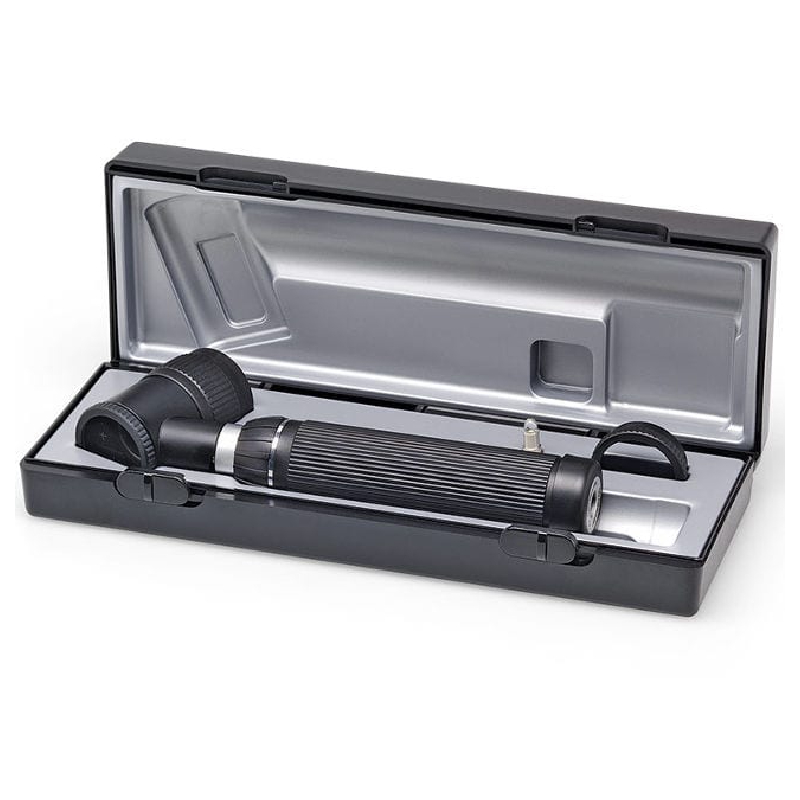
Riester ri-derma Dermatoscope for Skin Screening - Head Only
- Item: 3KM201376
Without anti-theft device
· No. 10550 XL 2.5 V
· No. 10551 XL 3.5 V
· No. 10577 LED 3.5 V
With anti-theft device
· No. 10551-301 XL 3.5 V
- Brand: Riester
Today skin cancer is a commonly occurring
disease, which can often be diagnosed and treated at an early stage. The
ri-derma dermatoscope offers physicians and specia-lists a reliable method of
dermatoscopic screening and early identification of malignant melanomas. Proven
epiluminescence microscopy allows melanocytic and non-melano-cytic structures
to be distinguished and enables precise observation of changes in skin pigmentation.
Without
anti-theft device
· No. 10550
XL 2.5 V
· No. 10551
XL 3.5 V
· No. 10577
LED 3.5 V
With
anti-theft device
· No. 10551-301
XL 3.5 V
• Illumination of the examination field with 3.5
V LED illumination and/or 2.5 V XL xenon illumination resembles natural
daylight.
• 10-fold magnification of the structure of the
skin with focussing, achromatic lens.
• Two different skin-friendly and sterilizable
contact plates are available:
- with scale
from 0 – 10 mm for exact measurement of pigmented skin lesions,
- without scale.
• Bulbs
can be quickly replaced at the base of the instrument head.
• Stable
casing of black chrome-plated metal, dustproof.
• Practical
eyeglass protection.
• Bayonet
fitting for quick and secure attachment of the instrument head to the handle.
• Large
selection of power sources: handy and stable handles, practical chargers and well-conceived
diagnostic stations.
• Large
selection of ri-derma sets at especially attractive prices.
• All of
the power sources can be combined with the high-quality Riester ri-derma line.
• Developed
and manufactured in Germany.
Specification
Customer Reviews
- No review for this product.


